Abstract
Gallstone ileus is a rare complication of cholelithiasis with high mortality rate related to advanced age and severe comorbidities of the patients, difficult diagnosis and subsequent delayed treatment. The most appropriate surgical strategy is still widely considered, especially the necessity of definitive management of the biliary pathology and the removal of the intestinal obstruction in one stage. We describe the case of 68-year-old female who presented with symptoms and signs for ileus-peritonitis. At the laparotomy the findings were: a fistula between gallbladder and duodenum with an impact gallstone in the duodenum (Bouveret?s syndrome) and distended small bowel to the distal part of the jejunum where a gallstone that caused the obstruction was identified. The patient underwent enterolithotomy, de-connection of the fistula, gallstone extraction from the duodenum and revision of the biliary ducts with subsequent pyloroplasty and sutures of the gallbladder.
Keywords :
Gallstone ileus
, Gallstone
, Biliary pathology
, Bouveret?s syndrome
Turkish Abstract
Safra taşı ileusu, hastaların ileri yaşına ve şiddetli ek hastalıklarına, zor teşhise ve sonrasında geciken tedaviye bağlı olarak yüksek ölüm oranına sahip ender bir kolelityaz komplikasyonudur. Safra patolojisinin definitif yönetim gerekliliği ve bağırsak obstrüksiyonunun bir aşamada çıkarılması da dahil olmak üzere, uygun cerrahi strateji halen geniş çapta değerlendirilmektedir. Bu olguda, İleus-karınzarı yanması şikayetleri ile başvuran 68 yaşındaki bir kadın hasta hakkında bilgiler verilmiştir. Laparotomi?de, safra kesesi ve duodenum arasında, safra taşının duodenum?a etkisiyle oluşan bir fistül (Bouveret sendromu) ve bir safra taşının obstrüksiyona sebep olduğu tanımlanan, jejunum?un distal bölgesine doğru şişmiş ince bağırsak bulguları tespit edildi. Hastaya entero-litotomi, fistül dekoneksiyonu, duodenum?dan safra taşının alınması ve buna müteakip piloroplasti ile safra yollarının revizyonu ve safra kesesi dikişi uygulandı.
Turkish Keywords :
, Safra taşı ileusu
, Safra taşı
, Safra patolojisi
, Bouveret sendromu
Introduction
Gallstone ileus is a rare complication of cholelithiasis, mostly in the elderly. The reported incidence is 1-3% of all types of intestinal obstructions and increases to 25% in patients older than 65 years. It is more frequent in women than in men with ratios raging from 4:1 to 16:1 1-4. High mortality rate is as high as 12-18% 1-5 related to advanced age, severe comorbidities, difficult diagnosis and subsequent delayed treatment. The most suitable strategy for emergency treatment of patients with gallstone ileus is still debated, especially the necessity of definitive management of the biliary pathology and the removal of the intestinal obstruction in one stage. The performance of enterolithotomy only saves the patient?s life but the risk of subsequent complications remains because of the available biliary-enteric fistula. They can be recurrent intestinal obstruction result from second gallstone, cholecystitis or recurrent cholangitis. On the other hand, one-staged enterolithotomy, cholecystectomy and closure of the fistula are related to prolonged operative time and higher operative risk, mortality and morbidity rates 4.
Case Report
A 68-years old woman was admitted to the Clinic urgently. One week before the hospitalization the complaints had started with colicky pain in right hypochondrial region and the epigastrium which progressively became permanent. The patient did not seek medical care. During the last 3 moths she had similar symptoms which were relieved for 2-3 days by taking antispasmodics. Gallbladder stones were diagnosed with ultrasound 10 years ago. Then she refused the suggested surgical treatment.
After 2-3 days nausea and vomiting started. The pain became permanent, fever to 380C with chills presented without visible jaundice. There were no disturbances in the intestinal passage. The patient reported gradual increase in complaints poorly responsive to antispasmodics. At the third day the pain decreased but fever remained permanent.
2 days later against the background of mild pain in upper right abdominal quadrant colicky pain in the whole abdomen accompanied by bowel rumbling and meteorism appeared. 3 days before the hospital admission the pain became permanent, without defecation and flatulence for this period. There were bloating and vomiting without bile impurity. The general condition of the patient at the time of hospital admission was impaired with manifested dehydration. Clinical examination revealed abdomen with bloating, diffuse tenderness and overt peritonism. Blumberg?s symptom was positive. The peristalsis was rumbling in overcoming nature. Digital rectal examination revealed that rectal ampulla was filled with normal colored stools. Biochemical and hematological investigations showed a white cell count of 22x109/ L, a hemoglobin of 103g/l, a red cell count of 3,5x10¹²/L, a total bilirubin of 56,3 mmol/l, a direct bilirubin of 34,7 mmol/l.
Abdominal X-rays demonstrated hydro-air levels originated from the small intestine in the upper abdomen and right abdominal half. The ultrasound imaging showed a contracted gallbladder with a thickened, in places double contoured wall, pneumobilia and gaseous distension of the intestines with a pathological cockade in small bowel. (Figure 1)
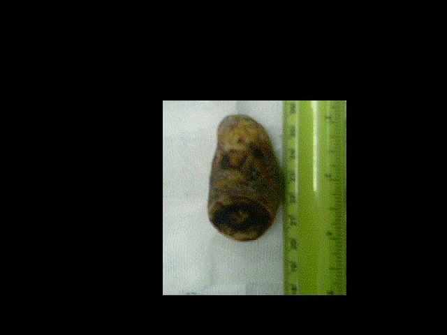
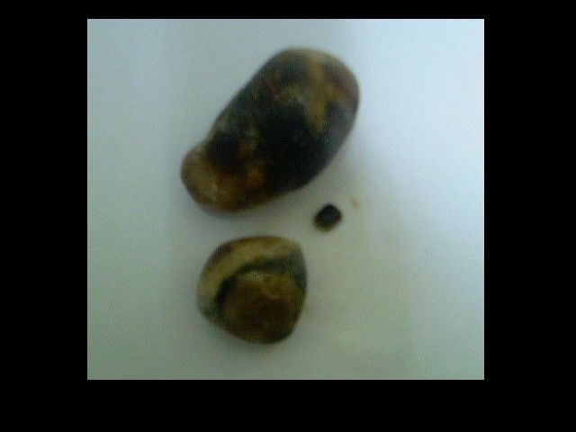
The condition is considered as ileus-peritonitis with decision for emergency surgical treatment. At the laparotomy the findings were: dilatation of the stomach, distended intestines to the distal part of the jejunum where a gallstone with a diameter of 25 mm that caused the obstruction was identified. The proximal small bowel was dilated (diameter of 15 mm) with spot areas on the serous layer and many deserosations. The gallbladder was covered by dense adhesions between surrounding tissues and omentum. After their liberation a fistula between gallbladder and duodenum with an impact gallstone in the duodenum were indentified (Bouveret?s syndrome). An enterolithotomy 15 cm proximally to the gallstone and sutures of the areas of deserosation were performed. Because of the impaired general condition of the patient it was considered that one-stage procedure was not indicated but the obstruction of the pylorus-duodenum required surgical management. So a longitudinal incision of the gallbladder, pylorus and duodenum with a fistulotomy was performed. The patient underwent de-connection of the fistula, gallstone extraction and revision of the biliary ducts with subsequent pyloroplasty and sutures of the gallbladder, too. (Figure 2, 3, 4)
Post operative recovery was unremarkable, the passage of the stools was recovered on day 3 and the patient was discharged home 13 d post operation.
We performed a follow-up two times in the first 30 days after discharge and on the 3-td moth after the operation. The control ultrasound showed a gallbladder with thicken wall, sludge, but without stones in the lumen. There was no pneumobilia. The patient had no serious complaints so a cholecystectomy was not considered.
Discussion
Cholelithiasis is a common disease but it is symptomatic in only 20-30% of the cases, most commonly presenting with biliary colic 1. The gallstone ileus is a rare complication of the disease. The gallstone ileus was first described and the term was coined by Bartolin in 1654 7. The major pathologic mechanism of gallstone ileus is the biliary-enteric fistula, which a gallstone migrates into the intestines thorough 4, 8. The stone may enter the intestines through a fistulous communication between the common bile duct and GI tract 1. The cases with ileus caused by migrated stone during papillotomy are rare. The stone size should be at least 2 cm to 2,5 cm in diameter to cause an obstruction 9. The most common site of gallstone impaction is the terminal ileum, but other localizations have also been described - jejunum, colon and even Meckel?s diverticulum 10,11.
Clinical presentation of gallstone ileus is nonspecific and more than 1/3 of the patients have no prior history of symptomatic disease which has been treated with antibiotics. The most common symptoms are nausea, vomiting and epigastric pain 1. Sighs typical to the gallstone ileus are colicky pain, bloating, disturbed bowel transit alternating with ?light? asymptomatic periods. A small portion of the patients may present with hematemesis result form duodenal erosions 1. The diagnosis of gallstone ileus is difficult. In over than 50% of cases, the diagnosis is made at laparotomy 4. The classic radiological Rigler?s triad which includes mechanical intestinal obstruction, pneumobilia and ectopic gallstone in the intestinal lumen has to be investigated 12. The presence of air in the gallbladder and pneumobilia are common findings in cases with gallstone ileus 13. The conventional abdominal X-rays usually are not sufficient because only 10% of gallstones contain enough calcium to be visualized radiographically 1. Abdominal ultrasound is a fast, harmless method but usually has not practical value, because the imaging is not clear for the presence of dilated bowel. Specific radiographical symptom is the ?Tambling obstruction?. Absence of stone in the gallbladder in patients with known cholelithiasis is also important sign which has to be taken into consideration. In recent years, CT significantly improves diagnosis but the percentage of patients who are diagnosed at laparotomy remains high 7.
The main aim of the treatment of gallstone ileus is to remove the intestinal obstruction. The most appropriate surgical strategy is still widely considered: enterolithotomy alone or enterolithotomy, cholecystectomy and fistula closure. The one-stage operation (enterolithotomy, cholecystectomy and fistula closure) excludes the opportunity for subsequent complications of the disease, but is related to higher mortality and morbidity rates 14. The two-stage procedure involves enterolithotomy as an emergency operation followed by second operation for cholecystectomy and closure of the fistula. It is recommended that the second procedure has to be performed 4-6 weeks after the initial operation. In the review of 1001 reported cases of gallstone ileus it was established that the mortality rate in one-stage procedure was 16,9% versus 11,7% in cases with 2-stage surgery 1. The recurrence rate of gallstone ileus after enterolithotomy alone was 5%. Prevention of these complications is the obligatory manual revision of the intestines for ?forgotten gallstones?. In 10% of the cases a relaparotomy may be required because of recurrent biliary symptoms 4. Enterolithotomy can be finished with ileostomy (in cases with severe ileus) or sutures of the intestinal wall. Small bowel resection with primary anastomosis is another therapeutic option in patients with gallstone ileus that has caused irreversible damage to the intestinal wall
Conclusion: Gallstone ileus remains a diagnostic and treatment problem with high morbidity and mortality rates. Patients commonly are admitted and surgically treated in emergency conditions of advanced bowel obstruction and severe general condition. The surgical strategy has to be individualized in relation to patient?s general condition and its aim must not be obligatory one-stage removal of the biliary pathology.
References
- Yakan S, et al. Gallstone ileus as an unexpected complication of cholelithiasis: diagnostic difficulties and treatment. Ulus Travma Acil Cerrahi Derg 2010;16 (4):344-8.
- Deitz DM, Standage BA, Pinson CW, McConnell DB, Krippaehne WW. Improving the outcome in gallstone ileus. Am J Surg 1986;151:572-6.
- Clavien PA, Richon J, Burgan S, Rohner A. Gallstone ileus. Br J Surg 1990;77:737-42.
- Reisner RM, Cohen JR. Gallstone ileus: a review of 1001 reported cases. Am Surg 1994;60:441-6.
- Balthazar EJ, Schechter LS. Gallstone ileus. The importance of contrast examinations in the roentgenographic diagnosis. Am J Roentgenol Radium Ther Nucl Med 1975;125:374-9.
- Beuran M, Ivanov I, Venter MD. Gallstone Ileus ? Clinical and therapeutic aspects. J Med Life 2010;3 (4): 365?71.
- Glenn F, Reed C, Grafe WR. Biliary enteric fistula. Surg Gynecol Obstet 1981;153:527-31.
- Rodríguez Hermosa JI, et al. Gallstone Ileus: results of analysis of a series of 40 patients. Gastroenterol Hepatol 2001;24:489-94.
- Schutte H, et al. Gallstone ileus. Hepatogastroenterology 1992;39:562-5.
- Nakamoto Y, Saga T, Fujishiro S, Washida M, Churiki M, Matsuda K. Gallstone ileus with impaction at the neck of a Meckel?s diverticulum. Br J Radiol 1998;71:1320-2.
- Beuran M, Venter MD, Ceauşu I.Ileusul biliar-aspecte clinicoterapeutice. Revista Medical? Na?ional? 1997; 1(4): 31-34.
- Balthazar EJ, Schechter LS. Air in gallbladder: a frequent finding in allstone ileus. AJR Am J Roentgenol 1978;131:219-22.
- Reisner RM, Cohen JR.Gallstone ileus. A review of 1001 cases. Am Surg 1994;60:441-6.
- Warshaw AL, Bartlett MK.Choice of operation for gallstone intestinal obstruction. Ann Surg 1966; 164: 1051-5.
|