Abstract
Intussusception is a rare disorder in adult patients. Diagnosis and treatment is challenging. Unique physical and radiographic examinations reveal valuable clues. We are presenting a case demonstrating all aspects of adult intussusception.
Keywords :
Adult
, Intussusception
, Diagnostic imaging
, Small intestine
Turkish Abstract
İntususepsiyon erişkinlerde nadir görülen bir durumdur. Tanı ve tedavisi zordur. Fiziksel ve radyolojik incelemeler önemli ipuçları verebilir. Bu çalışmada ince barsak lipomları nedeniyle gelişmiş olgumuzla birlikte erişkin intususepsiyon özelliklerini sunacağız.
Turkish Keywords :
, Erişkin
, İntususepsiyon
, Tanısal görüntü
, İnce barsak
Introduction
Intussusception is a disease of childhood, it is very uncommon in adults. It occurs about 5% in adults. Preoperative diagnosis is diffucult in adult patients with intussusception. It is %1-5 reason of intestinal obstruction in adults 1,2. In this case we are presenting an adult patient with intussuception as a result of intestional lipomas.
Case Report
A 50 year-old female patient was presented to our clinic with chronic abdominal pain which worsened in the last week. She was also vomiting after meals. Her complaints had started at least one year before her admission. Upper gastrointestinal imaging with barium was made and a colonoscopy was performed., which were all normal.
Physical examination revealed a mass in in the left upper abdomen and the patient felt pain with palpation. Bowel sounds were hyperactive. Abdominal X-ray revealed normal findings. Leucocyte count was within normal range (5900 x10³/uL [Range 3600-10000 x10³/uL]). A low sodium level of 134 mEq/L (136-147 mEq/L) was the only abnormality in blood biochemistry.
The abdominal tomography showed concentric alignment of jejunal segments into each other with mesenteric vessels like a whorl (Figure 1: Arrow shows the whorl).
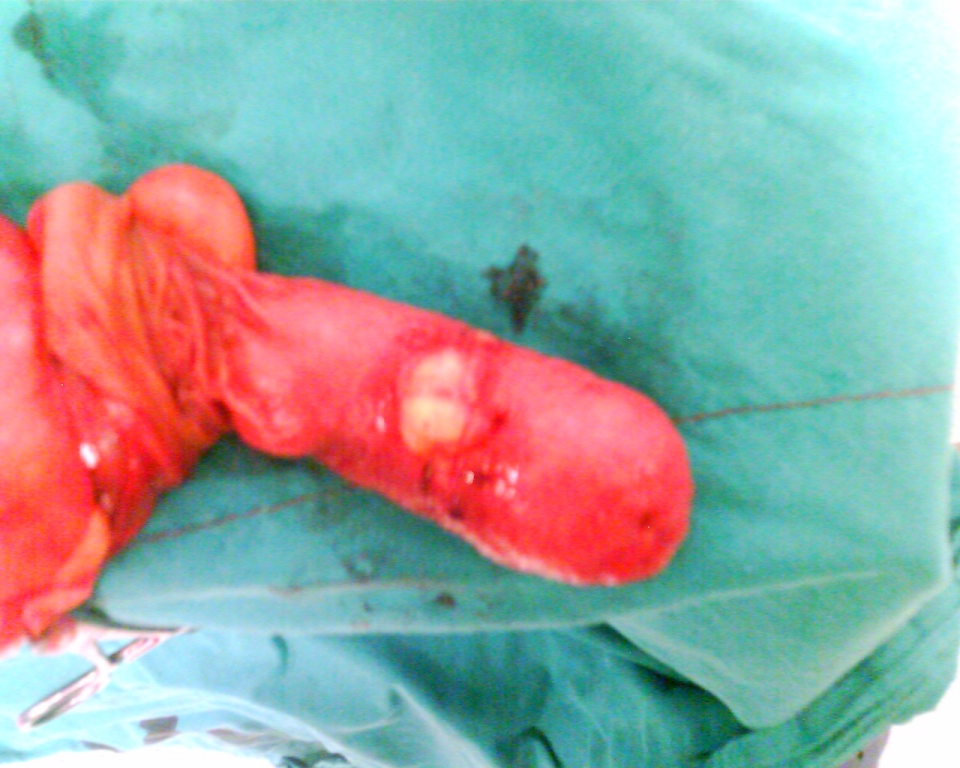
The patient was operated with the diagnosis of intussusception. The intussusceptum was a proximal jejunal segment about 40 cm from the ligament of Treitz. The distal jejunal segment was dilated. There was also a 4 cm long Meckel?s diverticulum, about 15 cm proximal to this segment. The diseased intestinal segment had several morphologic changes including neoplastic features, thus resection and primary anastomosis were performed. The pathology report revealed multiple lipomas growing into the lumen, the largest one being 8 cm in lenght (Figure 2).
Postoperative course was uneventful, and the patient was discharged one week after the operation.
Discussion
Intussusception occurs when a proximal segment of bowel (intussusceptum) telescopes or invaginates into the lumen of the adjacent distal segment (intussuscipiens).
Adult intussusception is rare. It is estimated that only 5% of all intussusception occurs in adults 1. It accounts for only 1% of intestinal obstructions 2. The presentation is non-specific which makes early diagnosis unlikely. The patients present with abdominal symptoms like nausea and vomiting. Ninety percent of adult intussusceptions occur in small or large intestines and 10% in stomach or surgically created stomas 3. Fewer than 20% of cases present acutely with complete bowel obstruction. On computerized tomography, a bowel-within-bowel configuration suggested by mesenteric fat and vessels compressed between the walls of small bowel is pathognomonic 4.
In the small intestine, there is a predominance of benign processes causing intussusception. These processes include polyps, hamartomas, lipomas, leiomyomas, neurofibromas, tuberculosis, inverted Meckel?s diverticulum and adhesions. Malignant alterations, primary or metastatic tumors, in small bowel invaginations are rare 5.
Lipomas are rare tumors of the small bowel comprising 16% of all benign tumors of the small bowel 5. Lipomas can occur in either in large or small bowel. They are usually submucosal and do not cause symptoms until they reach approximately 4 cm. Lipomas may cause chronic blood loss due to ulceration of the overlying mucosa in addition to intussusception 6.
Small intestinal lesions are more often benign and it is reasonable to attempt reduction first unless there are signs of bowel ischemia or a suspected malignancy to avoid resecting a long intestinal segment 7.
In cases of small bowel intussusception from adhesions, Meckel?s diverticulum, and benign polyps, a selective approach is feasible instead of en-bloc resection of the intestine. Adhesolysis, diverticulectomy or polypectomy are adequate treatments after reduction providing the bowel its viability 8.
References
- Martín-Lorenzo JG, et al. Intestinal invagination in adults: preoperative diagnosis and management. Int J Colorectal Dis 2004;19:68?72.
- Toso C, et al. Intussusception as a cause of bowel obstruction in adults. Swiss Med Wkly 2005; 135:87?90.
- Yalamarthi S, Smith RC. Adult intussusception: case reports and review of literature. Postgrad Med J 2005;81:174?177.
- Haas EM, et al. Adult intussusception. The American Journal of Surgery 2003;186:75?76.
- Gill SS, Heuman DM, Mihas AA. Small Intestinal Neoplasms. J Clin Gastroenterol 2001;33:267-282.
- Ross GJ, Amilineni V. Case 26: Jejunojejunal Intussusception Secondary to a Lipoma. Radiology 2000;216:727?730.
- Meshikhes AW, et al. Adult intussusception caused by a lipoma in the small bowel: report of a case. Surg Today 2000;35:161-165.
- Erkan N, et al. Intussusception in adults: an unusual and challenging condition for surgeons . Int J Colorectal Dis 2005;20:452?456.
|