Abstract
Multiseptate gallbladder is a rare congenital malformation. Although several asymptomatic cases have been described, patient usually present with right upper abdominal pain. We present a 42-year-old female patient's ultrasonography findings of multiseptate gallbladder.
Keywords :
Gallbladder
, Ultrasonography
, Abdominal pain
Turkish Abstract
Multisepta safra kesesi nadir bir konjenital malformasyondur. Genellikle asemptomatiktir. Sağ üst kadran ağrısı olabilir. Biz 42 yaşında multisepta safra kesesi olan kadın olguyu ultrasonografi bulguları ile sunacağız.
Turkish Keywords :
, Safra kesesi
, Ultrasonografi
, Abdominal ağrı
Introduction
Multiseptate gallbladder (MSG) is a rare congenital malformation of the gallbladder. Patients with MSG usually admits to the emergency services with differential symptoms such as right upper abdominal pain, nause and vomiting, and abdominal complaints 1.We present a female patient's ultrasonography (US) findings of MSG.
Case Report
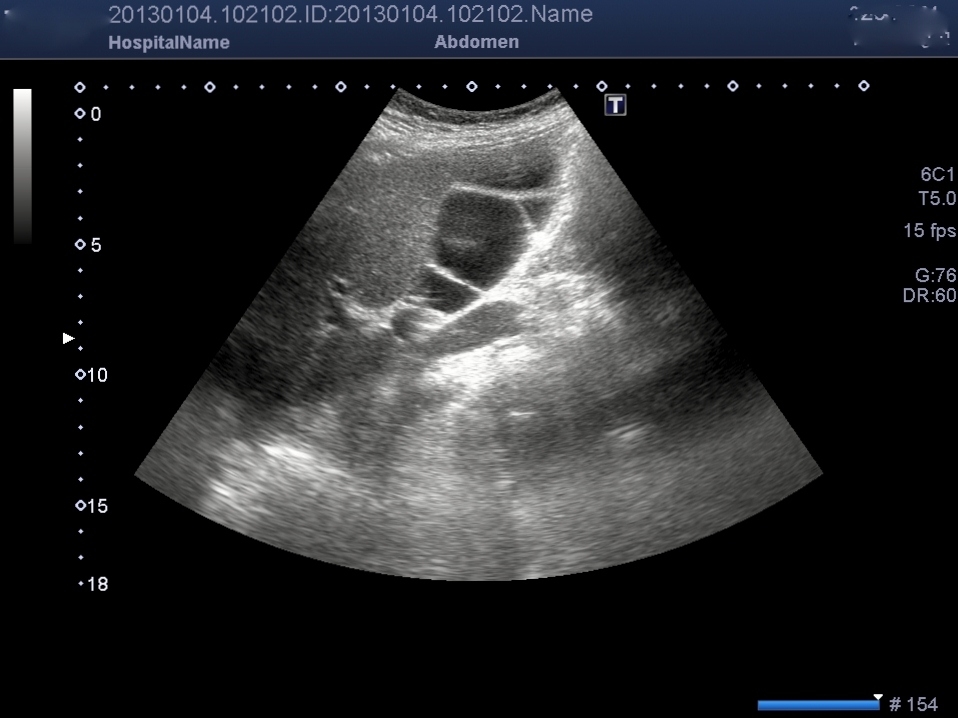
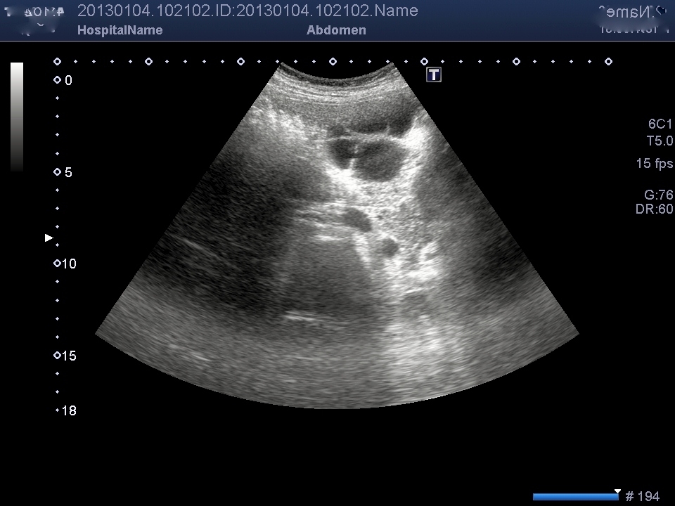
A 42-year-old female patient was admitted to our hospital with a complaint of recurrent right upper quadrant pain. The pain was associated with dyspepsia especially after fatty foods. Physical examination and laboratory tests (CBC, liver function tests, serum amylase, gamma glutamyl transpeptidase, and alkaline phosphatase levels, hydatid cyst tests) revealed no abnormality. An abdominal US was obtained with the suspect of gallstone. There were multiple linear echoes within the gallbladder dividing the lumen into compartments mimicking a honey-comb pattern. Multiple linear, fine echogenic bands without acoustic shadowing or septa crossing the lumen of the gallbladder (Figure 1-2).
The wall thickness of the gallbladder was normal, and there were no gallstones. Magnetic resonance cholangiopancreatography (MRCP) couldn?t performed because of claustrophobia. According to US findings, multiseptate gallbladder was diagnosed.
Discussion
MSG was first described in 1963 by Simon and Tandon 2. MSG is a rare congenital malformation of the gallbladder. It most likely results from incomplete vacuolization of the developing gallbladder bud or persistent ?wrinkling? of the gallbladder wall . Asymptomatic patients are very rare in literature , most patients present with long term abdominal symptoms such as right upper quadrant tenderness, recurrent abdominal pain, nause, vomiting, and gastrointestinal complaints 1. There is no reported association between uncomplicated. MSG and malignancy; however, there is a known link between biliary tract anomalies and cholangiocarcinoma. The incidence of malignancy in choledochal cyst is reported between 10-30%, and anomalous arrangement of the pancreaticobiliary duct is considered to be a high-risk factor for biliary tract malignancy 3. Anomalies of the gallbladder have been classified of a septum that divides the gallbladder lies longitudinally it is called bilobed gallbladder and when there is a transverse septum separating the fundus from the rest of the gallbladder it is called an hour-glass gallbladder. In patients with multiseptate gallbladder, US demonstrates multiple linear, fine echogenic bands without acoustic shadowing or septa crossing the lumen of the gallbladder, giving the organ a honeycomb appearance 4 . Septa are the reason of impaired motility of the gallbladder, and this causes a stasis in the bile flow, and it seems to be the reason of recurrent abdominal pain. US evaluation of the gallbladder is usually sufficient to diagnose MSG although other modalities such as computed tomography, MRCP and endoscopic retrograde cholangiography (ERCP) have been described to establish the diagnosis. The combination of US and MRCP is the most useful and the least invasive methods to diagnose MSG 1.On sonographic examination, desquamated gallbladder mucosa and the hyperplastic cholecystoses must be considered in the differential diagnosis.Desquamated gallbladder mucosa is seen as multiple linear echoes in the gallbladder lumen which do not arise from the wall of the gallbladder and the clinical setting is compatible with acute cholecystitis. The appearance of polypoid cholesterolosis and adenomyomatosis may mimic MSG, but there is no bridging of the gallbladder lumen by the cyst-like Rokitansky-Aschoff sinuses or polypoid bulbous echoes. A hydatid cyst should also be considered in the differential diagnosis, but the location and communication with the cystic duct and postprandial contraction of the gallbladder helped us to rule out this entity 5.
References
- Turgut K.,et.al. Diagnosis and Treatment of Multiseptate Gallbladder with Recurrent Abdominal Pain. Case Reports in Medicine (Internet). 2011, ( 2 page). Available from: http://www.hindawi.com/crim/medicine/2011/162853/
- Simon M., Tandon B.N. Multiseptate gallbladder: A case report. Radiology. 1963;80: 84?86
- Dylan W., et.al. Multiseptate Gallbladder in an Asymptomatic Child. Case Reports in GastrointestinalMedicine (Internet). 2011, ( 4 pages). Available from: http://www.hindawi.com/crim/gm/2011/470658/
- Nitin R. ,et.al. Septate gallbladder in the laparoscopic era. Journal of Minimal Access Surgery. 2008; 4-1:20-22
- Gülgün D., Gökhan D., Sadık T. Multiseptate gallbladder in a child with recurrent abdominal pain. Diagn Interv Radiol 2010; 16:306?307.
|