Abstract
Extracranial extension of intracranial meningiomas are rarely seen. This tumour had been reported in various anatomic sites.
In this paper, a rare case of intra- extracranial meningioma was reported.
Keywords :
meningioma
, intracranial
Turkish Abstract
İntrakraniyal menenjiomların ekstrakraniyal uzanımları nadir görülmektedir. Bu tümör çeşitli anatomik lokalizasyonlarda rapor edilmiştir.
Bu makalede nadir görülen intra-ekstra menenjiom olgusu sunuldu.
Turkish Keywords :
, menenjiom
, intrakraniyal
Introduction
Meningiomas are common tumours of the nervous system, representing 18% of primary intracranial tumors and 25% of primary intraspinal tumors1. Giant meningiomas with extracranial extension are unusual. In the literature a few cases of intracranial meningiomas with extracranial extension were reported2,3,4,5. Meningiomas are not usually considered in the differential diagnosis of head and neck swellings. We report here a meningioma with intracranial and extracranial components that presents with a temporal, maxillary region mass and ptosis.
Case Report
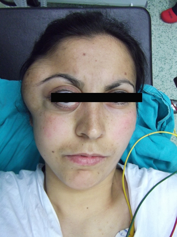
A 23 year old woman was admitted to our hospital with history of right temporal-maxillary swelling and proptosis of the right eye for one year. The proptosis was gradually getting worse and was associated with headache but no vomiting. Physical examination revealed a swelling of the right temporal region about 7x6 cms in diameter . The right eye was found to be markedly proptosed. There was no history of infection or ulceration of the mass (Figure 1).
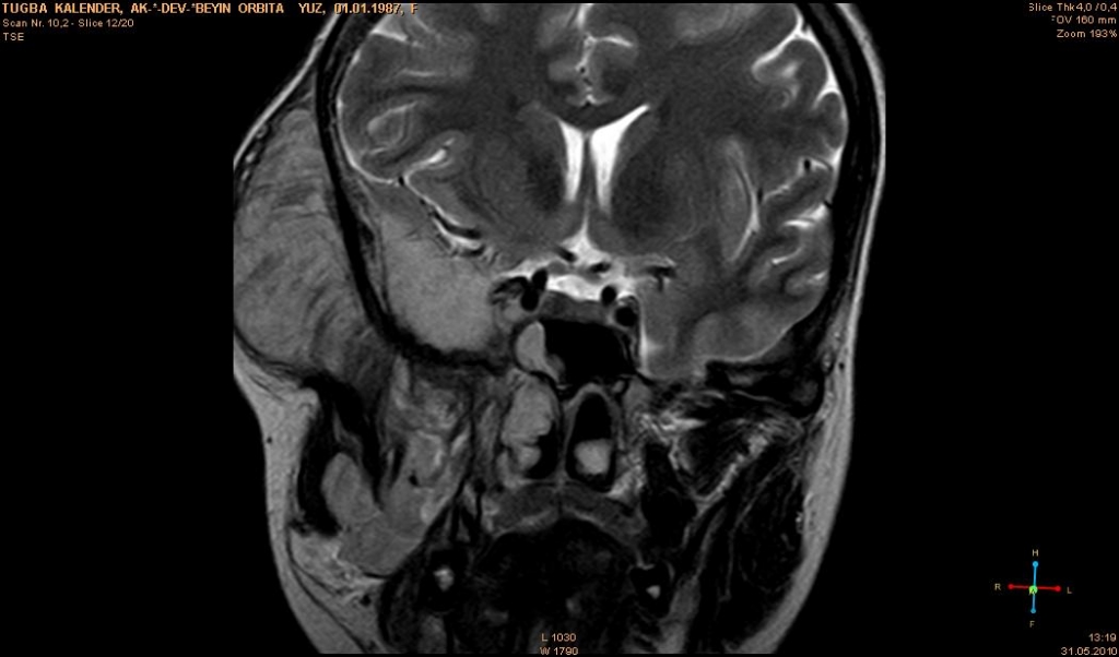
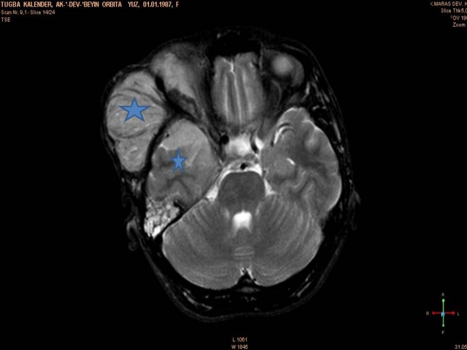
General appearence of patient was good. No other lesions or systemic involvement was found. Haemoglobin value was 4,9 gr/dl. Another laboratory test results were within normal limits. Magnetic resonance imaging (MRI) showed an extra-axial solitary mass lesion measured 11 cm × 7 cm within the right temporal lobe. There was extending mass into the right orbit, right maxillary sinus, and temporal, parapharyngeal, subgaleal region (Figure 2A-B).
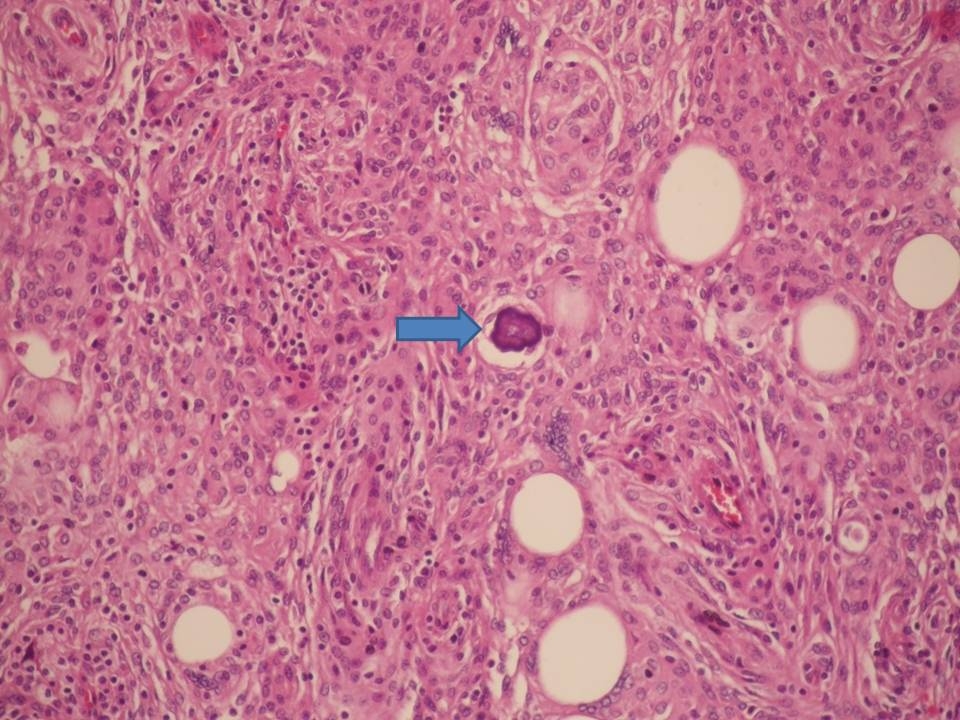
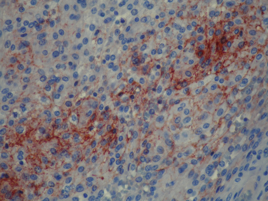
An incisional biopsy was performed in this region. Grossly two tissue masses were removed. The larger mass was firm, irregular circumscribed and grayish white-brown in colour, measured 9x8x1.5 cms. The small mass was measured 3x1x1 cms. Histologically, the cells were arranged in short fascicles and concentric whorls. The neoplastic cells had abundant, lightly eosinophilic cytoplasm, indistinct cytoplasmic borders and round or oval nuclei with finely dispersed chromatin and indistinct nucleoli. Nuclear clearing and lamellated calcospherules (psammoma bodies) were noted. Cellular atypia, necrosis, or increased mitotic activity was not detected. The tumor cells showed reactivity for Epithelial Membrane Antigen (EMA) and vimentin, but not for Glial fibrillary acidic protein (GFAP), Cytokeratin (CK), and S-100 protein (Figure 3A,B).
Meningotheliomatous (syncytial) meningioma corresponding to WHO grade I was considered as diagnosis on the basis of histopathological and immunohistochemical findings6. The patient was discharged for operation at faculty of medicine in a neighbour city.
Discussion
Meningiomas comprise the second largest group of primary brain tumors after gliomas7. Meningiomas are labeled only those neoplasms exhibiting morphologic or immunophenotypic evidence of an origin from meningothelial cells, specialised elements that populate the arachnoid membranes and cap the arachnoidal villi associated with intradural venous sinuses and their tributaries. Most meningiomas arise within the cranial cavity, are dura-based, and are found in the vicinity of the superior sagittal sinus, over the cerebral convexities or in contact with the falx cerebri6.
Extra-cranial meningioma is a tumour of rare occurrence. Only 2% of meningiomas occur extracranially as an estimation7. Four forms of the extra-cranial meningiomas have been suggested: 1. Primary intracranial meningioma with extracranial extension 2. Extracranial extensions of a meningioma arising in a neural foramen 3. Ectopic, without any connection either to a foramen of a cranial nerve or to intracranial structures 4. Metastases from intracranial meningiomas8.
Primary ectopic meningiomas have been reported in various anatomic sites in the ethmoid and maxillary sinus, submandibular region, foot, head and neck region, limp9,10,11,12,13.
Giant meningiomas with extracranial extension were reported unusual. These tumours present at sites such as the middle ear, in nasal cavity, in nasopharynx , in paranasal sinuses, in orbit as in our case, in oropharynx, in skin, in juxtaparotid14,6.
Bayar et al. and Dinc et al. reported two cases 29 and 54 years old patients of giant intra-extracranial meningioma at frontal and temporo-parietal lobe.But, they didn?t described histopathological subtype in their report2,3. Neeff et al. reported that patients with transitional subtype in the hypoglossal canal5. In another paper Arndt et al. reported intracranial meningothelial meningioma extension in left ethmoid and frontal sinuses15.
Preoperative diagnosis of intra-extracranial meningiomas may be difficult for the clinician. MRI and CT imaging is important for differential diagnosis of these tumours. A definitive diagnosis requires histopathological and immunohistochemical examination.
In conclusion, these tumors are unusual. It should be included in the differential diagnosis of soft tissue tumors. We evaluated a different location of meningioma extending to temporal, maxillary sinuses orbital cavity as a rare occasion.
References
- Rubinstein LJ. Tumors Of The Central Nervous System. Fascicle 6. Washington, DC: Armed Forces Institute of Pathology; 1972.
- Dinc C, Iplikcioglu AC, Cakabay M, Tufan A, Kosdere S.Intra-Extracranial Meningioma.Journal of Neurological Sciences (Turkish)2006; 23: 242-245.
- Bayar MA, Iplikcioglu C, Kokes F, Gokcek C.Intra-Extracranial Meningioma. Turkish Neurosurgery 1993;4: 170 - 172.
- Batsakis J G. Extracranial Meningiomas. Ann Otol Rhinol Laryngol. 1984;93:282?283.
- Neeff M, Baysal E, Homer J, Gillespie J, Ramsden R: Intracranial/Extracranial Meningioma Arising in the Hypoglossal Canal: Case Report.Skull Base. 2007 September; 17: 325?330.
- Rosai and Ackerman?s Surgical Pathology.Juan Rosai. eds Rosenblum MK, Bilbao JM, Ang LC Mosby .Ninth edition 2004;2:2564-2571.
- Friedman CG, Constantino P D. Primary Extracranial Meningiomas Of The Head And Neck. Laryngoscope 1990;100:41-48.
- Hoye SJ, Hoar CS, Murray JE. Extracranial Meningioma Presenting As A Tumor Of The Neck. Am J Surg 1960;100:486?489.
- Gokduman CA, Iplikcioglu AC, Kuzdere M, Bek S, Cosar M. Primary Meningioma Of The Paranasal Sinus. J Clin Neurosc 2005;12:832-834.
- Deshmukh S, Rokade V, Pathak G, Nemade S, Ashturkar A: Primary Extra-Cranial Meningioma İn The Right Submandibular Region Of An 18-Year-Old Woman:A Case Report. Journal Of Medical Case Reports 2011; 5:271.
- Tomaru U, Hasegawa T, Hasegawa F, Kito M, Hirose T, Shimoda T: Primary Extracranial Meningioma Of The Foot: A Case Report. Jpn J Clin Oncol 2000; 30:313-317.
- Hameed A, Gokden M, Hanna EY: Fine-Needle Aspiration Cytology Of A Primary Ectopic Meningioma. Diagn Cytopathol 2002; 26:297-300.
- Tuncay IC, GulerUO, VuralC, AkgunRC, DemirorsH, KuruI :Primary Extracranial Meningioma Of The Lower Limb.Joint Diseases And Related Surgery,2011;22:114-117.
- Thompson LDR, Gyure KA. Extracranial Sinonasal Tract Meningiomas. Am J Surg Pathol 2000;24:640-650.
- Arndt S, Thorsten Wiech, Mader I, Aschendorff A, Maier W.Rare Extracranial Localization Of Primary Intracranial Neoplasm: Case Report. Diagnostic Pathology 2008;3:14.
Information Presentation
This article was accepted at the 20. International Congress of Pathology as poster presentation.
|