Abstract
Branchial anomalies are the most common congenital neck pathologies in the lateral aspect of the neck. They are mostly asymptomatic until become infected. Branchial cleft cyst(BCC) is often become enlarged after upper respiratory tract infection attacks and become distinct as a painless, fluctuant, neck mass. A 15-yearold girl presented to the hospital with a left-sided neck mass and a 29-year-old women was admitted to the hospital presented with a swelling on the right side of the neck. They were operated and the pathologic specimens were reported as the branchial cleft cyst. Congenital neck masses can be misdiagnosed as lymphadenitis. The branchial cleft cyst should be kept in mind in the patient presenting with a lateral neck mass.
Keywords :
Second branchial cleft cyst
, Neck mass
, USG
, MRI
Turkish Abstract
Brankiyal anomaliler boynun yan tarafında en sık görülen doğumsal boyun patolojileridir. Enfekte olana kadar çoğunlukla asemptomatiktirler. Brankial yarık kisti (BCC) sıklıkla üst solunum yolu enfeksiyonu ataklarından sonra büyür ve ağrısız, fluktant bir boyun kitlesi olarak ortaya çıkar. Boyun sol tarafında kitle şikayeti ile hastaneye başvuran 15 yaşındaki kız çocuğu, 29 yaşında kadın boynun sağ tarafında şişlik şikayeti ile hastaneye ile başvurdu. Ameliyat edildiler ve patoloji spesmenleri brankial kleft kisti olarak rapor edildi. Konjenital boyun kitleleri lenfadenit olarak yanlış tanı alabilirler. Yan boyun kitlesi ile başvuran hastalarda brankial kleft kisti akılda tutulmalıdır.
Turkish Keywords :
, İkinci brankial kleft kisti
, Boyun kitlesi
, USG
, MRG
Introduction
Branchial cleft cysts are congenital lesions caused by anomalous development of the branchial arches. Branchial cleft anomalies can be in the form of cysts, sinuses, and fistulas and develop between four and seventh weeks of embryonic life. The second branchial cleft cyst is the most common type (%95)1. They can be situated at the anterior border of the sternocleidomastoid muscle, between the mandibular angle and the clavicle 2.
The branchial cleft cyst(BCC) is mostly asymptomatic until becomes infected. They are often become enlarged after upper respiratory tract infection attacks and become distinct as a painless, fluctuant, neck mass 3. They rarely become malignant 4. Although BCCs are congenital and might be found at birth, most are not detected until the first or second decade of life. The diagnosis is usually made by ultrasound, computed tomography, and fine-needle aspiration biopsy. But definitive diagnosis and treatment of BCC are made by surgical resection and pathologic examination 1. This case report describes 15 and a 29-year-old female with swelling on the lateral aspect of the neck.
Case Report
Case Report 1
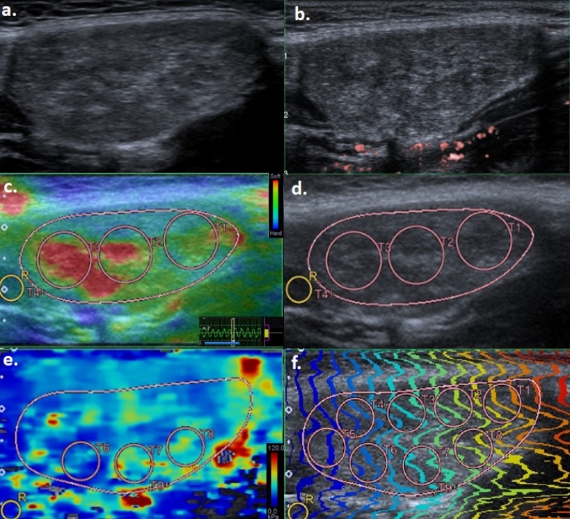
A 15-year-old girl was admitted to hospital complaining about swelling of the left side of the neck that developed over the past 3 years and enlarged for the last 1 month. A lesion which shows solid appearance in B-mode, no vascularity signals in Superb microvascular imaging(SMI), soft in strain elastography(SE)(Strain index (SI) value: 0.12 ) and intermediate shear-wave velocities in shear wave elastography (SWE)( 41,7 kPa, 3,68 m/s) observed in ultrasonography(US) (figüre 1).
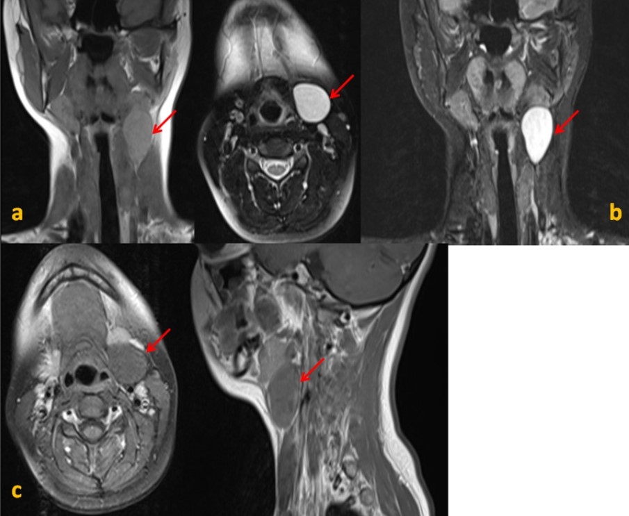
In contrast-enhanced magnetic resonance imaging(MRI) of the neck, posterior to the left submandibular gland, anterior to the sternocleidomastoid muscle (SCM), 37x27x26 mm in size, mild hyperintense on T1- weighted images, hyperintense signal on T2- weighted images (figüre 2a), non-enhancing lesion was found after IV contrast injection (figüre 2b). The lesion was excised and reported as a branchial cleft cyst on histopathological examination.
A 29-year-old women was admitted to the hospital presented with a swelling on the right side of the neck that gradually increased in size in the last 9 months. In physical examination, a mobile mass, 4x3 cm in size, sensitive to touch, slightly hyperemic was observed in the right cervical region. A hypoechoic lesion with dimensions of 37x25 mm, evaluated as lymphadenopathy, was detected in the neck ultrasonography. In contrast-enhanced MRI of the neck, posterior to the right submandibular gland, deep to the SCM, 50x28 mm in size, hyperintense on T1- weighted images (figüre 3a), containing hypointense areas on T2- weighted images, showing fluid-fluid level (figüre 3b), showing diffusion restriction in places in DWI (figüre 3c), showing peripheral contrast enhancement after intravenous contrast injection, well-defined cystic lesion was observed.
Case report 2
A 17x16 mm lymphadenopathy was seen anterior to the lesion. The lesion was excised and reported as a branchial cleft cyst on histopathological examination.
Discussion
Branchial cleft cysts are congenital lesions caused by anomalous development of the branchial apparatus 5. BCC is a common cause of soft tissue swelling in the lateral aspect of the neck in late childhood and early adulthood. It generally occurs unilaterally 6. In 1955, Proctor described four classes of branchial cleft cyst 2. Type I BCC anomalies are of ectodermal origin. It is a duplication anomaly of the external ear. It can be seen deep in the platysma, anterior to the SCM. Type II BCC is the most common type (%95). The second BCC is located inferior to the mandibular angle and anterior to the SCM muscle, abutting the internal carotid artery and adherent to the internal jugular vein. The sinus tract of the cyst passes through the deep structures of the neck and opens to the tonsillar fossa. Rarely, malignancies arising from the epithelium layer of the cyst may occur. The third BCC is located more inferior to the neck and anterior to the SCM muscle compared to the second, extending medially between the carotid bifurcation and the lateral wall of the pharynx. The fourth BCC is very rare, it lies in the pharyngeal mucosal space; lined with columnar epithelium 2.
Clinically, patients are usually asymptomatic. When it becomes symptomatic, presents as a slow-growing, solitary, painless compressible swelling in the anterior triangle of the neck 3. Sometimes it can grow fast and this acute change in the size of the mass is often caused by an upper respiratory tract infection such as pharyngitis, otitis media, or dental infection 7.
The first choice of radiological method in the diagnosis of BCCs is US. It is easily accessible and there is no ionizing radiation. Ultrasonography is usually sufficient for the diagnosis of uncomplicated BCCs. Computed Tomography and MRI is used in the diagnosis and localization of complicated BCCs and the identification of the relation to surrounding tissues. Diagnosis depends on clinical suspicion and knowledge of the typical location. The definitive diagnosis of branchial cleft anomalies is made by surgical excision and pathologic examination 1,2.
The BCC can be misdiagnosed with other space-occupying lesions in the neck. The differential diagnosis of branchial cleft anomalies includes cervical lymphadenitis, thyroglossal duct cyst, dermoid cyst, metastatic cystic lymph nodes of squamous cell carcinoma, HIV-related lymphadenopathy, papillary thyroid carcinoma metastasis, neck abscess, lymphangioma, tuberculosis 2,8. Infected congenital neck masses can be confused with lymphadenitis. Congenital anomalies may be the underlying cause in the diagnosis of recurrent or not regressing lymphadenitis despite appropriate treatment 9. Solitary cystic metastases of head and neck cancers can clinically mimic BCC. Neck lymph node metastasis of tonsillar and anterior tonsillar plica cancers may have cystic content. Among cystic lymph node metastases, the rate of originating from the tonsil is between 33-50%. Also, papillary thyroid cancer can cause cystic neck lymph node metastasis in the same way 10.
The possibility of metastatic carcinoma should be considered, especially in older adult patients even in the absence of clinical symptoms 11. Therefore, preoperative fine-needle aspiration cytology(FNAC) helps plan the surgical strategy and exclude malignant disease 12.
BCC management starts with infection control if there is an infection. Surgical excision may be considered in terms of controlling recurrent infections and preventing complications of these infections 4. Although rare, the most important reason for surgery is the malignant potential of BCC. Other treatment methods are aspiration, sclerotherapy, and incision and drainage. However, these methods can cause recurrence and increase the risk of secondary infection 5.
Conclusion
The branchial cleft cyst should be considered in the lateral neck swelling in young adults and pre-diagnosed lymphadenitis that recur or does not regress despite treatment.
References
- Adams A, et al. Branchial cleft anomalies: a pictorial review of embryological development and spectrum of imaging findings. Insights Imaging. 2016;7(1):69-76.
- Bocchialini G, et al. Unusually rapid development of a lateral neck mass: Diagnosis and treatment of a branchial cleft cyst. A case report. Int J Surg Case Rep. 2017;41:383-6.
- KÖNte E, et al. Branchial Cleft Cyst in the Differential Diagnosis of Recurrent Lymphadenitis. Türkiye Çocuk Hast Derg. 2018; 3: 212-14.
- Lee DH, et al. Clinical Study of Second Branchial Cleft Anomalies. J Craniofac Surg. 2018;29(6):e557-e60.
- Bagchi A, et al. Branchial cleft cysts: a pictorial review. Pol J Radiol. 2018;83:e204-e9.
- Valentino M, Quiligotti C, Carone L. Branchial cleft cyst. J Ultrasound. 2013;16(1):17-20.
- Şahin K, et al. A Rare Case of A Mass on the Neck that Changes its Dimensions When Weeping. Istanbul Medical Journal. 2017;18:97-9.
- Tansuker H, et al. Branchial cleft cyst: A rare diagnosis in a 84-year old patient. A case report. Turkish Archives of Otolaryngology. 2011;49:22-4.
- H. D. Tansuker, et al. Turk Otolarengoloji Arflivi / Turkish Archives of Otolaryngology. 2011; 49:22-4.
- Könte EK, et al. Türkiye Çocuk Hast Derg/Turkish J Pediatr Dis / 2018; 3: 212-14.
- Sellami M, Ghorbel A. Branchial cleft cyst: a case report. Pan Afr Med J. 2017;26:102.
- Al Sukhun J, El Naggar M. Unusual Presentation of a Large Multilocular Second Branchial Cleft Cyst. J Craniofac Surg. 2019;30(6):1772-3.
|