Abstract
In female patients, pelvic masses are frequently originate from the reproductive organs. In addition, pelvic masses may arise from the gastrointestinal system, urinary system, neurogenic and soft tissues and metastases. In this paper, we present a 75-year-old female patient with a pelvic mass.
Keywords :
Neurogenic mass
, pelvic organ
, misdiagnosis
Turkish Abstract
Kadın hastalarda, pelvik kitleler sıklıkla reprodiktif organlardan kaynaklanır. Bununla birlikte, gastrointestinal sistem, üriner sistem, nörojenik ve yumuşak dokular, metastazlardan pelvik kitleler ortaya çıkabilir. Bu yazıda pelvik kitlesi olan 75 yaşında bir kadın hasta sunuldu.
Turkish Keywords :
, Nörojenik kitle
, pelvik organ
, yanlış tanı
Introduction
In women, gynecologic masses may constitute an important part of the pelvic masses, but masses that do not originate from the gynecological organs may also be seen1. Differentiating gynecologic masses from gynecologic mass imitators is important for the treatment plan.
Case Report
A 75-year-old female patient complained of pelvic pain for the past 1 year and an increased pain of a 1-week duration. This was the first time he was admitted to hospital for his complaints. Therefore, he had no medical records of previous physical examination and laboratory tests. Ultrasonography (US) examination performed at the external center revealed a heterogeneous mass with 8x7 cm in size in the right adnexal region and the patient was informed and pelvic magnetic resonance imaging (MRI) examination was performed.
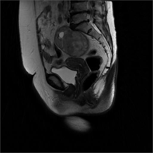
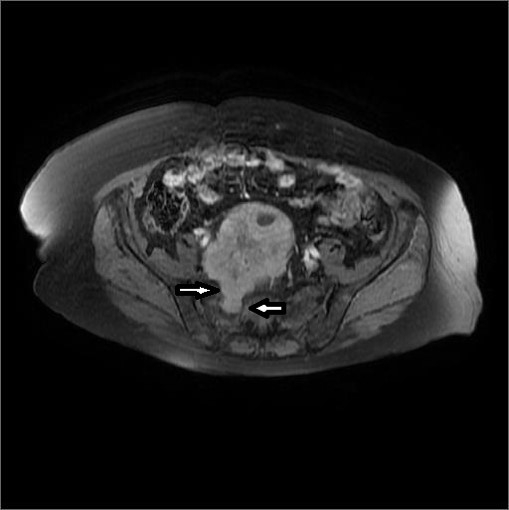
The pelvic MRI examination (Optima 360, GE Helthcare, Milwaukee, USA) showed a heterogeneous, hypointense hyperintense signals on T1W images, heterogeneous, iso-hyperintense signals on T1W images, markedly graded after IV contrast agent injection. There was a focal component extending from the sacral level to the right neural foramen tongue (Figure 1-2).
The mass was not related to the uterus or the ovaries. Radiologically, it was considered as a ?neurogenic tumor?. Histopathological examination with true-cut biopsy revealed a "benign neurogenic tumor".
Discussion
In female patients, the majority of pelvic masses arise from the uterus and ovarian masses.
There are lesions that can be confused with primary ovarian masses due to pelvic localization 2. It may be difficult to distinguish the pelvic masses located between the anatomic compartments on US scans. In this case, computerized tomography (CT) and especially MRI may provide reliable and useful information in detecting the localization of the pelvic masses. Therefore, MRI is a useful diagnostic tool for the diagnosis and for planning the operation3. In the literature, pelvic abscess, cyst hydatid, malignant lymphoma, mesenteric cysts, retroperitoneal mucinous tumors, appendix mucosal, postoperative or posttraumatic hematomas, gossypiboma, neurogenic tumors and bone pelvic tumors / soft tissue lesions are reported as gynecologic mass imitators 2-4. There are cases of non-gynecological origin lesions that are misjudged with gynecologic origin masses, without any further imaging in the literature 5. This creates problems with patient management during or after the operation.
In the pelvic region; neurogenic tumors such as schwannomas, neurofibromas and paragangliomas can be observed. Schwannomas, are generally well-defined tumors located in the pelvic presacral area.
Schwannomas usually appear as enhancing lesions with calcification and cystic degeneration, less than 5 cm on CT scans. MRI features are not specific and are hypointense on T1W images, hyperintense on T2W images. Neurofibromas can be observed especially in cases of neurofibromatosis and almost everywhere in the body. On CT and MRI scans they may appear with large sizes and infiltration . Paragangliomas are tumors that originate from the paraganglionic system and can be observed in different localizations. On CT and MRI scans, paragangliomas are seen as more vascular lesions compared to well-defined ones. CT and MRI are important as they may show the origin of a lesion. However, US, CT and MRI generally do not provide benefit in determining pathologic subtypes of neurogenic tumors 1,3.
In the present case; the pelvic mass which could not be distinguished from gynecologic pathologies with US, was defined as neurogenic tumor by demonstrating the sacral foraminal extension on MRI scans and the diagnosis was confirmed histopathologically.
In conclusion; CT and MRI should be used in addition to the US to detect the location of a lesion and internal structure characteristics in terms of reasoning and appropriate patient management. A radiologist should keep in mind that majority of pelvic region masses in women are of gynecologic origin, but non-gynecologic lesions may also be observed.
References
- Levine CD, et al. Benign extraovarian mimics of ovarian cancer. Distinction with imaging studies. Clin Imaging. 1997;21:350-8.
- Ozat M, ve ark. Extraovarian conditions mimicking ovarian cancer: a single center experience of 15 years. Arch Gynecol Obstet. 2011;284:713-9.
- Karaosmanoglu AD, Ozmen MN. Jinekoloji dışı pelvik kitlelerde görüntüleme. Trd Sem 2015; 3:83-91.
- Ateser G, ve ark. Adneksiyal kitleyi taklit eden pelvik retroperitoneal kitle: Bir schwannoma olgusu. İstanbul Tıp Dergisi. 2006;2:40-2.
- Ibraheim M, Ikomi A, Khan F. A pelvic retroperitoneal schwannoma mimicking an ovarian dermoid cyst in pregnancy. J Obstet Gynaecol. 2005;25:620-1.
Information Presentation
37th National Radiology Congress, 1-6 November, 2016, Antalya, Turkey
|