Abstract
Even though lipoma is the most common soft tissue tumor, pure lipoma primary to uterus is very rare. As fat tissue is not native to the uterus, various theories of histogenesis have been proposed such as metaplasia of the embryonic fat cells, proliferation of perivascular fat cells. Clinical symptoms and physical signs are similar to those found in leiomyomas, except that they mostly affect postmenopausal women. In this article we report the radiological findings of a lipoma which was incidentally diagnosed with ultrasound and tomography in a women who admitted to the hospital with pelvic pain and we review the literature.
Keywords :
Uterine
, Lipoma
Turkish Abstract
Lipom en sık görülen yumuşak doku kitlesi olduğu halde uterin yerleşim oldukça nadirdir. Uterusun yapısında yağ dokusu bulunmadığından uterin lipomun gelişiminde embriyonik yağ dokunun metaplazisi, perivasküler yağ dokunun proliferasyonu gibi teoriler öne sürülmektedir. Leyomyomlara benzer semptom verir ve postmenopozal dönemde daha sık görülürler. Bu olgu sunumunda pelvik ağrı şikayeti ile hastaneye başvuran kadın hastada yapılan abdominal ultrasonografide ve kontrastsız abdominal tomografide insidental olarak saptanan uterin lipoma ait radyolojik bulguları literatürü gözden geçirerek sunmayı amaçladık.
Turkish Keywords :
, Uterin
, Lipom
Introduction
Even though lipoma is the most common soft tissue tumor, pure lipoma primary to uterus is very rare 1. Occasionally the clinical presentation or macroscopic appearance may mimic a sarcoma and create diagnostic confusion 2,3. Clinical symptoms and physical signs are similar to those found in leiomyomas, except that they mostly affect postmenopausal women 4. The histogenesis of these lesions is still unclear 5.
Case Report
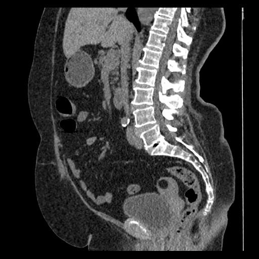
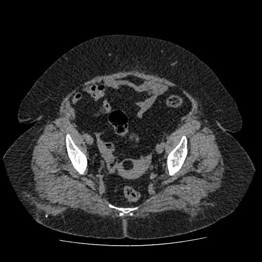
32 year-old female patient who admitted to the hospital with pelvic pain. Incidentally a hyperechoic uterine mass was detected in abdominal ultrasonography (Figure 1). In non-enhanced abdominal computed tomography it was a hypodense lesion with pure fatty attenuation (Figure 2,3).
Discussion
The histogenesis of these lipomatous tumors in the uterine wall continues to be an enigma 3. As fat tissue is not native to the uterus, various theories of histogenesis have been proposed. These include misplaced embryonic fat cells, metaplasia of the muscle or connective tissue cells into the fat cells, lipocytic differentiation of specific primitive connective tissue cells, proliferation of perivascular fat cells accompanying the blood vessels into the uterus, inclusion of the fat cells into the uterine wall during surgery or fatty infiltration or degeneration of the connective tissue 6. Uterine lipomas were first described by Lobstein in 1816.7 Most uterine lipomas are found in postmenopausal women between the ages of 50 and 70 years 8. They often present with symptoms similar to uterine fibroids including chronic pelvic discomfort, heaviness, or pressure. Approximately 50% have associated uterine bleeding 9. Seraphini et al, performed ultrasonographic analyses in 11 lipomatous lesions of the uterine corpus which they observed as avascular masses with well-defined hyperechogenic margins 10. The highly echogenic central region corresponds to the fatty tissue with the hypoechoic rim thought to correspond to the myometrium incorporated into the growing tumor.8,11 Because of the fatty nature of the tumor, CT can be diagnostic when the tumor?s location within the uterus is apparent and those mixed lesions with predominantly Ieiomyomatous elements may appear identical to uterine fibroids.8 The uterin myolipoma can be a mixed (fat and solid tissue) uterine mass in CT scan, however, diagnosis may be difficult if the mass is exophytic or pedunculated.12 On T2-weighted MR images, fat content within the tumour can be confirmed because of evident chemical shift artifact.13On MRI, the lipoma has high signal intensity on T1 and T2 weighted images. The fat content is clearly demonstrated with fat suppression techniques.14 In a study of lipomas compared to liposarcomas all tumors without a recognizable nonadipose component were benign lipomas and when specific findings are encountered upon MRI examination, the physician should consider a pure uterine lipoma.15
Although the diagnosis of a uterine lipoma was made following surgery, current imaging techniques enable us make the diagnosis without surgery.5 When specific findings are encountered upon MRI examination, the physician should consider a pure uterine lipoma.15
References
- Garg A, et al. Pure lipoma of uterus: A case report with review of literature. J Sci Soc. 2013;40:114-5.
- JacobsJDS, Cohen H, Johnson JS. Lipoleiomyomas of the uterus. Am Clin Pathol. 1965;44:45-51.
- Gupta RK, Hunter RE. Lipoma of the uterus: review of literature with views of histogenesis. Obstet Gynecol. 1964;24:255-7.
- Al-Maghrabi JA, Sait KH, Lingawi SS. Uterine lipoma. Saudi Med J. 2004;25:1492-4. PubMed PMID: 15494831.
- Dilek TU, et al. Uterine lipoma and coincidental cervical cancer: a case report. Int J Gynecol Cancer. 2006;16(1):445-7.
- Dharkar DD, Kraft JR, Gangaharan D. Uterine lipoma. Arch Pathol Lab Med. 1981;105:43-5.
- Gaspar IA. Lipoma of the uterus: a study and demonstration of the histogenesis of two cases. J Med Soc NJ. 1980:77(3): 179-82.
- Jacobs JE, Markowitz SK. CT diagnosis of uterine lipoma. AJR. 1988;150(6):1335-6.
- Krenning RA, DeGoey WB. Uterine lipomas. Review of the literature. Exp Obstet Gynecol. 1983:10(2-3):91-4.
- Serafini G, et al. Lipomatous tumors of the uterus: ultrasonographic findings in 11 cases. J Ultrasound Med. 1996;15(3):195?9.
- Houser LM, Carrasco CH, Sheehan CA. Lipomatous tumour of the uterus: radiographic and ultrasonic appearance. Br J Radiol. 1979:52:992-3.
- Vilallonga R, et al. Lipoma of the uterine corpus: exceptional eventuality combined with an ovarian thecoma. Case Rep Med. 2009; Article ID 340603. doi: 10.1155/2009/340603. Epub 2009 May 26.
- Mylona S, et al. Uterine lipoleiomyoma: transvaginal ultrasound and computed tomography findings of an unusual entity. European Clinics in Obstetrics and Gynaecology. 2008;3(3-4):135?7.
- Coumbaras M, et al. Uterine lipoma: MRI features with pathologic correlation. Abdom Imaging. 2005; 30: 239?41.
- Ohguri T, et al. Differential diagnosis of benign peripheral lipoma from well-differentiated liposarcoma on MR imaging: is comparison of margins and internal characteristics useful? AJR. 2003;180(6):1689-94.
|