Abstract
Cholesterol granuloma is a granulomatous inflammation in reaction to the cholesterol. It can develop in air cells of the temporal bone. This condition can occur in middle ear cavity, mastoid cells and petrous apex. It is described as one of the most common benign lesion of the petrous apex. Herein, we report a case of cholesterol granuloma in petrous apex with the clinical and radiological findings.
Keywords :
Cholesterol granuloma
, Petrous apex
, CT
, MRI
Turkish Abstract
Kolesterol granülomu, kolesterolle reaksiyona giren granülomatöz bir enflamasyondur. Temporal kemiğin hava hücrelerinde gelişebilir. Bu durum orta kulak boşluğunda, mastoid hücrelerde ve petröz apekste ortaya çıkabilir. Petroz apeksin en sık görülen iyi huylu lezyonlarından biri olarak tanımlanır. Burada petröz apekste kolesterol granülomu olgusunu klinik ve radyolojik bulgularla sunuyoruz.
Turkish Keywords :
, Kolesterol granülomu
, Petrous apeks
, BT
, MRG
Introduction
Cholesterol granuloma (CG) is a granulomatous inflammation in reaction to the cholesterol crystals and red cell extravasation. It can develop in air cells of the temporal bone with inadequate drainage. This condition can occur in middle ear cavity, mastoid cells and petrous apex 1, 2. It is described as the most common benign pathological lesion of the petrous apex 1. Herein, we report a case of cholesterol granuloma in petrous apex with the clinical and radiological findings.
Case Report
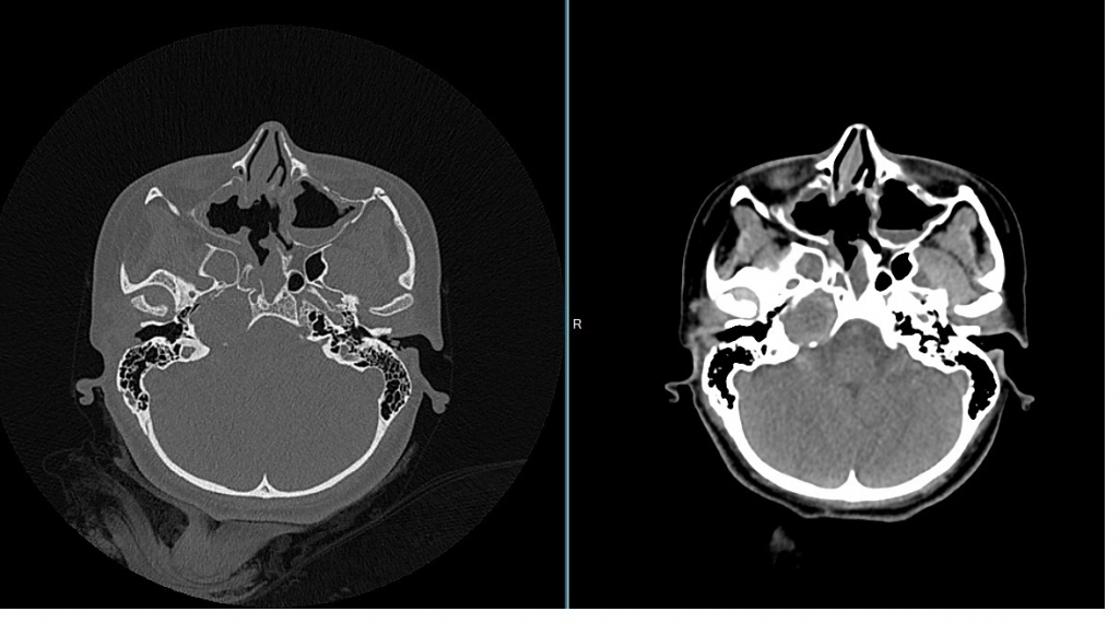
A 15-year-old woman had undergone CT scan after traumatic incident. Computed tomography (CT) scan revealed an ovoid, isodens mass of about 2 cm in the petrous apex. The lesion expanded petrous apex and smoothly eroded the surrounding bone (Figüre 1).
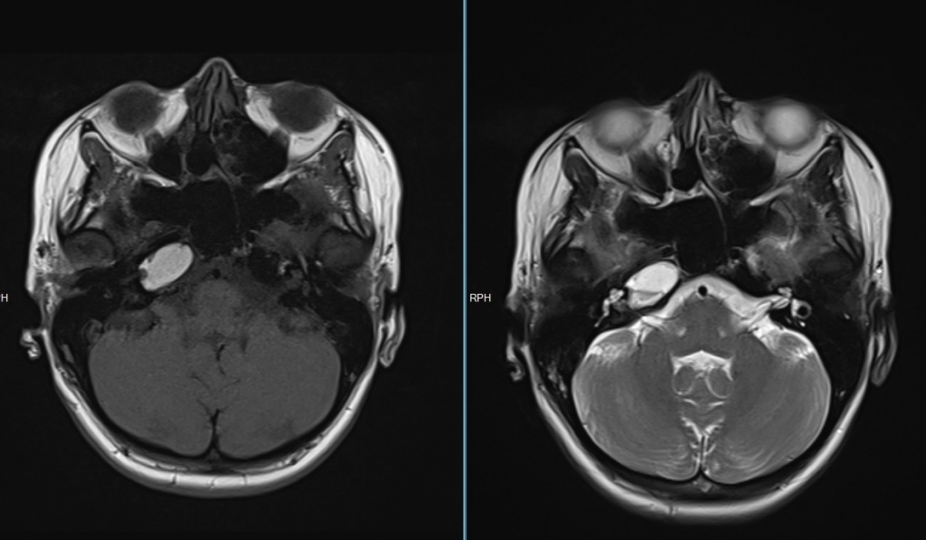
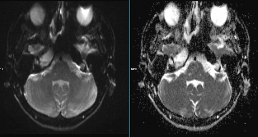
Due to these findings, the patient underwent contrast-enhanced temporal magnetic resonance imaging (MRI) scan. The lesion showed homogen hyperintensity on T1-weighted images and heterogen hyperintensity on T2-weight images without diffusion restriction (Figüre 2a,b).
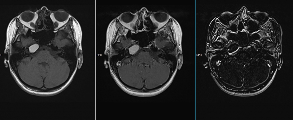
After contrast administration, it showed faint peripheral enhancement (Figüre 3). Because our patient has no symptoms, surgical intervention was warranted. The lesion haven’t showed any change during five-year follow-up.
Discussion
Cholesterol granuloma in the external and middle ear cavity was first described by Manasse in 1894 3. It is an osteolytic lesion with a chronic granulomatous reaction. It is not limited to the temporal bone, but can occur in other locations too: central nervous system, frontal bone, orbital cavity, paranasal sinus, infratemporal fossa, thyroid gland, mediastinum, ovary and peritoneum 4,5.
The pathogenesis of petrous apex CG has been related to two hypotheses: One, the obstruction-vacuum-theory where eustachian tube dysfunction is thought to be the underlying abnormality and the exposed marrow theory in which hyperplastic mucosa invades the underlying bone and exposes bone marrow, which in turn bleeds 6.
For diagnosis, MRI findings of CG are unique. It shows high signal on both T1-weighted and T2-weighted images, which remains high after fat suppression due to chronic hemorrhage. Contrast administration is not required for diagnosis. Epidermoid and dermoid cysts show similar CT findings, but loss signal on fat supression images and epidermoid cysts demonstrate restricted diffusion.
Petrous apex CG can present as hearing loss, dizziness, tinnitus. If symptomatic, surgical treatment is required. Because our patient has no symptoms, surgical intervention was warranted. The lesion haven’t showed any change during five-year follow-up.
Informed Consent
hastadan
References
- Pace A, Iannella G, Riminucci M, Corsi A, Magliulo G. Tympano-Mastoid Cholesterol Granuloma: Case Report and Review of the Literature. Clin Med Insights Case Rep. 2020; 13: 1179547620958728.
- Terao T, Onoue H, Hashimoto T, Ishibashi T, Kogure T, Abe T. Cholesterol granuloma in the petrous apex: case report and review. Acta Neurochir (Wien). 2001; 143: 947-52.
- Manasse P. Ueber Granulationsgeschwulste mit Fremdkoerperresenzellen. Virch Arch. 1984; 136: 245.
- Ugga L, Stilo S, Napolitano P, et al. Orbitofrontal cholesterol granuloma: case report and review of the literature. Quant Imaging Med Surg. 2017;7:373-77.
- Rinaldo, A, Ferlito, A, Cureoglu, S, Devaney, KO, Schachern, PA, Paparella, MM. Cholesterol granuloma of the temporal bone: a pathologic designation or a clinical diagnosis? Acta Otolaryngol. 2005;125: 86-90.
- Graham, MD, Kemink, JL, Latack, JT, Kartush, JM. The giant cholesterol cyst of the petrous apex: a distinct clinical entity. Laryngoscope. 1985;95:1401-6.
Information Presentation
30. TNRD Yıllık Toplantısında Poster Olarak Kabul Edildi
|