Abstract
Bronchial anatomy cross-sectional imaging and reconstruction images are shown on three-dimensional images with the appropriate computerized tomographic technique. Unlike numerous variations of the lobar or segmental bronchial subsections, abnormal bronchi from the trachea or main bronchi are rare. Among these, accessory cardiac bronchus and ?tracheal? bronchus are among the bronchial abnormalities. Sometimes congenital bronchial abnormalities may not be diagnosed unless there is any complication until advanced ages. However, knowing and understanding this variation is important for diagnosis, bronchoscopy, surgery and intubation.
Keywords :
Trachea
, Bronchus
, variation
, anomaly
, Computed tomography
Turkish Abstract
Bronşiyal anatomi kesit görüntüleme ve rekonstrüksiyon görüntüleri bilgisayarlı tomografik teknikle üç boyutlu görüntülerle gösterilmiştir. Lober veya segmental bronşiyal alt bölümlerin sayısız varyasyonunun aksine, trakea veya ana bronşlardan anormal bronşlar nadirdir. Bunlar arasında aksesuar kardiyak bronş ve ?trakeal? bronş, bronşiyal anormallikler arasındadır. Bazen ileri yaşlara kadar herhangi bir komplikasyon olmadıkça konjenital bronşiyal anormallikler teşhis edilemeyebilir. Bununla birlikte, bu varyasyonu bilmek ve anlamak; tanı, bronkoskopi, cerrahi ve entübasyon için önemlidir.
Turkish Keywords :
, Trakea
, Bronş
, varyasyon
, anomali
, Bilgisayarlı tomografi
Introduction
Accessory tracheal bronchus is an anatomical variant that is directly attached to the trachea wall from a bronchus supracarinal level. The incidence is estimated to be approximately 1% (in the range of 0.1-2%) and there is a pronounced right-hand dominance 1. We aimed to present a case of tracheal bronchus with computed tomography images.
Case Report
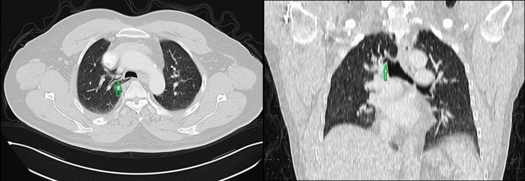
A 57-year-old male patient underwent computed tomography of the thorax during follow-up for solitary pulnomer nudule; Tracheal bronchus was observed in the right upper lobe segment at the supracarinal level of the trachea (Figure1a-b).
Discussion
It was first described by Sandifort in 17852. Developmental bronchial anomalies usually occur in infancy or early childhood, but may not show symptoms in some people. Anomaly can be detected incidentally in computed tomography scans such as our case. Tracheal bronchus may cause recurrent infection and respiratory distress in children. Rarely, during the application of anesthesia or treatment of respiratory failure, unintentional tracheal bronchi can be intubated. Obstruction, atelectasis and post-obstructive pneumonia may cause respiratory failure.
As a result; Tracheal bronchus is a rare congenital development which causes focal aeration, pneumonia or emphysema in a small number of cases. In children with recurrent pulmonary infection or in patients who will undergo surgery for any reason, it should be recognized before surgery, intubation and kept in mind by radiologists and clinicians to guide treatment.
References
- Shih FC, Lee WJ, Lin HJ. Tracheal bronchus. CMAJ. 2009; 180 (7): 783.
- Ghaye B, Szapiro D, Fanchamps JM et al. Congenital bronchial anomalies were reviewed. The radiographs taken. 21 (1): 105-19.
|