Abstract
Kaposiform hemangioendothelioma is a rare locally aggressive vascular tumor of intermediate malignancy. Altough it is usually presented as a skin lesion, involvement of so many unusal sites have been reported.Herein we report a case of kaposiform hemangioendothelioma in a 39 year old women that is presented with a superficial cutanous lesion on the forearm.
We are reporting this case because of its rarity in adults and also because of difficulties in differentiaL diagnosis with other vascular tumors that have similiar histological features.
Keywords :
Kaposiform hemangioendothelioma
, Cutaneous
, Adult
Turkish Abstract
Kaposiform hemanjioendotelyoma nadir görülen lokal agresif bir tümördür. Genellikle kutanöz yerleşimli olmakla birlikte, çok çeşitli lokalizasyonlarda izlenmektedir. Burada 39 yaşında ön kolunda yüzeyel yerleşimli kutanöz bir kitle ile karşımıza çıkan kaposiform hemanjioendotelyoma olgusu sunulmaktadır. Bu vaka bu tümörün erişkinlerde oldukça nadir görülmesi ve benzer özellik taşıyan diğer vaskuler tümörler ile ayırıcı tanıda sıkıntı yaratması açısından önem arz etmektedir.
Turkish Keywords :
, Kaposiform hemanjioendotelyoma
, Kutanöz
, Erişkin hasta
Introduction
Kaposiform hemangioendothelioma is a rare locally agressive vascular endothelial neoplasm that occurs nearly exclusively during childhood. Altough it is usually identified in infancy and first decade, adult cases have been reported recently 1. It is typically presented as a superficial or deep soft tissue mass of extremities especially proximal arms and legs as an ill- defined, red-to-purple induretad plaque 2. The diagnosis is based on histologic examination that is characterized by infiltrating nodules and sheets of spindle cells and similar histologic features to Kaposi?s sarcoma that can lead to misdiagnose. It grows rapidly with focal extension into the adjacent skin, soft tissue and bone.
Case Report
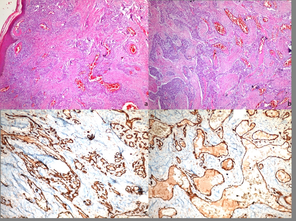
A 39 year old women with a subcutanous lesion; 0,5 x 0,3 x0,4 cm diameter in her forearm. In microscopic examination with H&E stained sections of this lesion there is a tumor that is subcutanous located and inflitrative through to dermis (Figure 1a). Tumor is comprised of slit- like vessels that are lined by spindle shaped endothelial cells without distinct pleomorphism (Figure 1b). Mitotic activity is rare and there is no extravasated erythrocytes in tumor. In immunohistochemical examination of tumor; there is positive staining with CD31, CD34 and SMA (Figure 1c-d). Ki67 proliferative index of tumor cells is < %5. There is negative staining with HHV8.
Discussion
Kaposiform hemangioendothelioma is first described by Zukerberg and colleagues in 1993 as a distinctive lesion of childhood that has features common to both capillary hemangioma and Kaposi sarcoma 3.
Kaposiform hemangioendothelioma rarely occurs in adults older than 21 years 4. Based on a literature search; Fernandez et al determined fewer than 20 adult patients with kaposiform hemangioendothelioma since it was first described by Zukerberg and colleagues in 1993 2. Several features of kaposiform hemangioendothelioma in adults are different from those in children. Despite the equal sex distribution in children, there is a male predominance with a ratio of 80% in adults 4. Also in contrast to large tumors (diameter > 5 cm) in children, adult-onset kaposiform hemangioendotheliomas are small 5.
Although, 75% of kaposiform hemangioendothelioma presented with cutaneous involvement, numerous anatomic locations have been reported; cervicofacial region (sinuses, external and internal auditory canals, larynx, thymus, thyroid, eyelids), trunk, mediastinum and retroperitoneum 6. Also, unusual sites such as the deltoid muscle, spleen, uterine cervix, thoracic spine, breast, regional lymph node metastases and even pulmonary involvement have been documented 2,7.
There is no certain information about tumorigenesis yet it is suggested that tumorigenesis of kaposiform hemangioendothelioma can develop in the embryonic period 1.
Kaposiform hemangioendothelioma is associated with lymphatic vessel proliferation and Kasabach-Merritt phenomenon (KMP) which presents with severe trombocytopenia, consumptive coagulopathy, and microangiopathic anemia 1. This phenomenon is not often observed in adults because of its small size 5.
The most important differential considerations in this disease are Kaposi sarcoma, cellular hemangioma of infancy, solitary juvenile hemangioma and multiple hemangiomas 8. And also angiosarcoma and acquired tufted hemangioma should be taken in consideration for differential diagnosis especially in adult patients. Immunohistochemical stains are useful in substantiating a definite diagnosis. Positive staining for CD34, CD31, ERG and SMA indicates vascular origin. Also vascular endothelial growth factor receptor-3 and monoclonal antibody D2-40 are suggestive of lymphatic differentiation 2. GLUT-1 can help to differentiate kaposiform hemangioendothelioma from juvenile hemangioma by totally lack of immunoreactivity. In addition to this; HHV8 staining can distinguish between Kaposi?s sarcoma which is related to HHV8 transcripts in tumorigenesis 1.
Because of its invasive growth pattern, no-self healing tendency for kaposiform hemangioendothelioma is classified as intermediate (borderline) malignancy 1. Despite its local aggressiveness, it does not have any metastatic potential. In addition to this, death from kaposiform hemangioendothelioma has resulted from severe coagulopathy 8. The treatment of this tumor is based on wide local resection. If it is associated with KMP, adjuvant medical theraphy is required 1.
In conclusion, although its rarity in adults, kaposiform hemangioendothelioma should be taken in consideration for differential diagnosis with other cutaneous vascular tumors especially with Kaposi?s sarcoma and angiosarcoma because of the different treatment modalities.
References
- Liu Q, et al. Clinicopathological features of Kaposiform hemangioendothelioma. Int J Clin Exp Pathol. 2015;8(10):13711-8.
- Kim MG, Choi YS, Park SJ, Chong SM. Kaposiform hemangioendothelioma of the breast in an adult female. Clin Breast Cancer. 2011;11(2):135-7.
- Zukerberg LR, Nickoloff BJ, Weiss SW. Kaposiform hemangioendothelioma of infancy and childhood. An aggressive neoplasm associated with Kasabach-Merritt syndrome and lymphangiomatosis. Am J Surg Pathol. 1993;17(4):321-8.
- Lyons LL, et al. Kaposiform hemangioendothelioma: a study of 33 cases emphasizing its pathologic,immunophenotypic, and biologic uniqueness from juvenile hemangioma. Am J Surg Pathol. 2004;28(5):559-68.
- Wu CH, et al. Expansile kaposiform hemangioendothelioma deformed thoracic cage in an adult. Ann Thorac Surg. 2013;96(5):1854-7.
- Croteau SE, et al. 3rd. Kaposiform hemangioendothelioma: atypical features and risks of Kasabach-Merritt phenomenon in 107 referrals. J Pediatr. 2013;162(1):142-7.
- Azma R, et al. Multifocal kaposiform hemangioendothelioma of soft tissue with bilateral pulmonary involvement in an adolescent. Korean J Pediatr. 2014;57(11):500-4.
- Mac-Moune Lai F, et al. Kaposiform hemangioendothelioma: five patients with cutaneous lesion and long follow-up. Mod Pathol. 2001;14(11):1087-92.
Information Presentation
Olgu 2-6 Kasım 2016 tarihindeki 26. Ulusal Patoloji ve 7. Ulusal Sitoloji Kongresi? nde ve 25-29 Eylül 2016 tarihindeki 28. Avrupa Patoloji Kongresi? nde sunulmak üzere kabul edilmiştir.
|