Abstract
Gas containing renal stone is an unusual and rare entity of kidney and can be life-threatening as an evidence of emhysematous pyelonephritis in certain clinical settings. Its early recognition is fundamental for a favorable outcome. Disease is usually seen in females with diabetes mellitus, with or without obstructive uropathy. We present a fatal case of gas containing renal stones in a patient without diabetes but with hemiplegia.
Keywords :
Emphysematous pyelonephritis
, Renal stone
Turkish Abstract
Gaz içeren renal taşlar böbreğin nadir görülen bir olgusu olup, bazı klinik durumlarda amfizematöz pyelonefritin belirteci olarak hayatı tehdit edebilirler. Olumlu bir sonuç için erken tanı esastır. Hastalık genellikle obstruktif üropatisi olan yada olmayan diyabetes mellitus hastalarında izlenir. Biz burada diyabetes mellitusu olmayan hemiplejik bir hastada ölümcül seyreden gaz içeren renal taş olgusunu sunduk.
Turkish Keywords :
, Amfizematöz pyelonefrit
, Renal taş
Introduction
The presence of gas in the upper urinary tract, along with renal calculi, has been described in the setting of recent urinary tract instrumentation, emphysematous renal infections, and gastrointestinal and pulmonary fistulas involving the urinary tract 1. Gas containing renal stone is a rare condition with clinical manifestations mainly presented as hematuria, proteinuria or flank pain. They usually occur in people with diabetes mellitus. Although most of the gas containing renal stones have been previously reported in patients with a history of diabetes, we present a case of gas containing renal stones in a patient with hemiplegia who is also prone to urinary tract infection.
Case Report
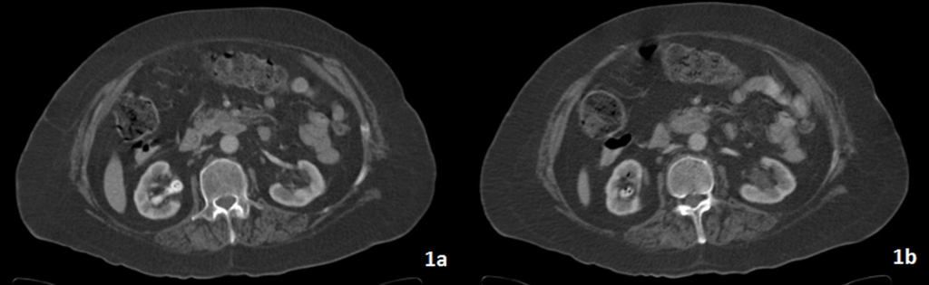
A 60 year-old female patient who has small sized lip wound for over 5 years was hospitalized for plastic surgery. Her medical history included epilepsy, hypertension and hemiplegia. Other physical examination findings and all laboratory values were normal. At the time of the pre-operative evaluation, her body temperature was increased. In order to investigate the source of fever, contrast enhanced abdomen computed tomography (CT) examination was performed. On CT, there were hyperdens stones in right renal calices, which include gas bubbles (Figure 1a-b).
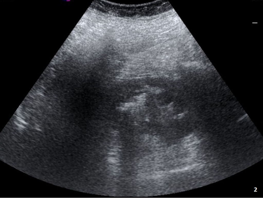
Meticulous evaluation of the patient revealed no other source of fever (such as abscess or infection of any kind). Ultrasound examination revealed hyperechoic stones, however failed to demonstrate the gas bubbles within the stones (Figure 2).
Based on CT findings, urinary tract infection caused by gas-forming microorganisms was suggested. When urine culture was obtained E.coli was isolated as the cause of this infection. Surprisingly on the following days her condition progressively deteriorated and E.coli was also isolated from blood culture despite initiation of prompt and appropriate antibiotic treatment. Subsequently septic shock occurred and renal failure developed. GFR value was 17ml/sec and creatinin level was 4,5 mg/dL. One week after her admission to hospital, despite all the medical treatment the patient died from urosepsis, septic shock and renal failure.
Discussion
Renal stones containing gas are very rare. The exact etiology of this unusual finding has not yet been definitively determined but may be an early, or variant, manifestation of emphysematous pyelonephritis (EPN). EPN is a life threatening condition with gas-producing, and necrotizing infection involving the renal parenchyma, collecting system, or perirenal tissue with a mortality rate that can be as high as %70 2. Most cases occur with uncontrolled diabetes mellitus. But in this case our patient did not have diabetes. On the other hand she had right sided hemiplegia which might raise the tendency to urinary tract infection 3. Although the main offending pathogen is E. coli as in this particular case; micro-organisims such as Proteus, Pseudomonas, Klebsiella, Enterobacter, and Candida have also been reported as causative organisms 2.
Abdominal X-ray can show gas within the renal shadow. But the low sensitivity of plain x-ray makes it inadequate for diagnosis and follow-up. The value of ultrasound for the evaluation of such renal stones is differently appreciated by various authors. Nevertheless, stone with gas, which mainly contains fibrotic tissue, proteins and necrotic cells like all other renal stones due to infection, isn't as dense as calcified urinary tract stones 4. Therefore, it is stated that posterior acoustic shadowing might not be so clearly seen in these patients which might help for differentiation of renal stone type. However, in our case we were able to demonstrate acoustic shadowing due to renal stones with gas. CT scan has been advocated as the most useful modality for diagnosing this entity. There are distinct characteristic features on CT that we can use to reveal the location, shape and internal structure of a stone besides no contrast material is required 1. It is fairly easy to depict gas within a renal stone. For localized emphysematous pyelonephritis, the success rate of antibiotic treatment, with or without percutaneous drainage, has improved substantially in recent years 5. Even though our patient had only small air bubbles in renal stone with no air in renal parenchyma or other parts of urinary tract, she died because of sepsis and shock despite aggressive antibiotic therapy. This might be due to micro-organisms settled within the stone itself and might be resistant to antibiotic treatment. Her clinical condition worsened with the advance of acute renal failure. Contrast material which was used during CT exam may have facilitated the progression of renal failure. Although Wan et al reported that the mortality rate was greater in patients with serum creatinine level >1,4 mg/dl and thrombocytopenia (platelets<60,000/mm³), our patient's all laboratory results were normal 2.
It has been stated that the mortality rate in patients managed medically is greater than that in patients managed surgically, while combined treatment has produced a survival rate of more than 90% 2,4. It has also been claimed that immediate nephrectomy is necessary, since delayed operation may only increase mortality5.Conversely, several patients have been treated successfully with percutaneous drainage, control of diabetes, and institution of broad-spectrum antibacterial therapy.
Conclusion
In conclusion, gas containing renal stone is an unusual life threatening entity. It is regarded as a type of EPN and should be considered in patients who are prone to urinary tract infection such as diabetes mellitus. When gas bubbles are found within the renal stones, one should keep in mind that this type of infection might be severely resilient to antibiotic treatment.
References
- Huang JJ, Tseng CC. Emphysematous pyelonephritis: clinicoradiological classification, management, prognosis, and pathogenesis. Arch Int Med. 2000;160:797-805.
- Wan YL, et al. Predictors of outcome in emphysematous pyelonephritis. J Urol. 1998;159:369-73.
- Langhorne P, et al. Medical complications after stroke: A multicenter study. Stroke. 2000;31:1223-9.
- Bani-Hani AH, Segura JW, Leroy AJ. Urinary matrix calculi: our experience at a single institution. J Urol. 2005;173:120-3.
- Best CD, et al. Clinical and radiological findings in patients with gas forming renal abscess treated conservatively. J Urol. 1999;162:1273-6.
Information Presentation
Kasım 2013'de Antalya'da düzenlenen 34. Ulusal Radyoloji Kongresi'nde poster olarak sunulmuştur.
|