Abstract
We present a case of sinolith in the maxillary sinus. She was a 38-year-old woman who complained of foul-smelling nasal discharge, heaviness on the left cheek, postnasal drip, halitosis and intermittent cough. Computed tomography (CT) showed a smooth-margined oval stone in the left maxillary sinus. The etiology, radiological features, differential diagnosis and treatment of the disease were discussed in the lights of literature.
Keywords :
Sinolith
, Antrolith
, Maxillary sinüs
, Rhinosinusitis
Turkish Abstract
Bu yazıda kötü kokulu burun akıntısı, sol yanakta ağırlık hissi, geniz akıntısı, ağız kokusu ve aralıklı öksürük şikayetleri olan 38 yaşında kadın hasta vesilesiyle maksiller sinüste sinolit olgusu sunduk. Bilgisayarlı tomografi görüntülemesinde sol maksiller sinüs içinde düzgün kenarlı oval bir taş görüldü. Hastalığın etyolojisi, radyolojik özellikleri, ayırıcı tanısı ve tedavisi literatür verileri eşliğinde tartışıldı.
Turkish Keywords :
, Sinolit
, Antrolit
, Maksiller sinüs
, Rinosinüzit
Introduction
Sinolith means a stone in the paranasal sinuses . The occurance of sinolith is very rare. Maxillary sinus is the most commonly involved sinus and then ethmoid, frontal and sphenoid sinüs 1. Most antroliths are small and asymptomatic. Larger ones may present with nasal purulant secretion, foul-smelling discharge, postnasal dripping, chronic cough and recurren and drug resistant rhinosinusitis. Long-standing infection, poor sinus aeration and drainage seem to be the most important predisposing factors of the stone formation 2.
Case Report
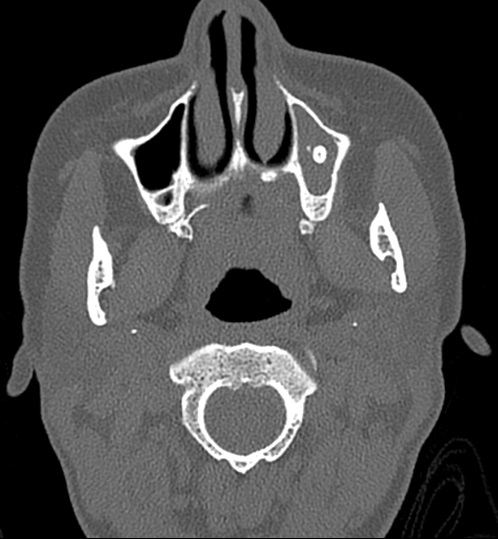
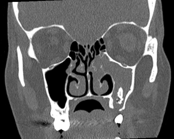
We present a case of sinolith in the maxillary sinus. She was a 38 year-old-woman who complained of foul-smelling nasal discharge, heaviness on the left cheek, postnasal drip, halitosis and intermittent cough. Patient have had a dental operation three years ago and there is an oro-antral fistula at the left second molar tooth region. Paranasal computed tomography (CT) revealed an opasification in the left maxillary sinus and inflammatory process around it (Figure 1 and 2).
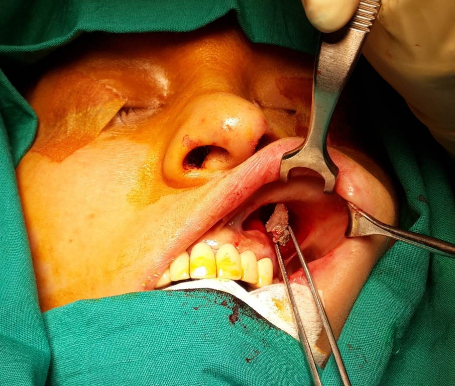
During surgery, we exposed and removed the stone with endoscopic and transoral Caldwell-Luc technique together, then repaired oro-antral fistula with lateral mucosal flep and bones of maxillary sinus anterior wall obtained from transoral Caldwell-Luc technique. (Figure 3 and 4).
The histopathological exam results as sinolithiasis in maxillary sinüs.
Discussion
Sinoliths are considered by many authors to be dystrophic calcificationor ossification caused by chronic inflammation of the paranasal sinus, also other pathologic conditions, including neoplastic disease, trauma and after radiotherapy 3.
Surgical removal of the stone is usually performed together with appropriate treatment of the coexisting sinus disease. In the literature, most of extractions from the outer approach have been reported 4. However, an endoscopic approach is satisfactory for the treatment of ethmoid sinoliths and maxillary sinus antroliths 5. Patients with antrolith may be asymptomatic and may be incidentally discovered on routine radiological examination 6. The usual clinical features in symptomatic patients are facial pain, nasal obstruction, epistaxis, purulent or blood-stained discharge, foul-smelling postnasal drip.
However, dacryocystitis, otorrhoea, anosmia, palatal perforation, and septal perforation have been reported in the literatüre 7. Radiographically, a dense, irregular yet well-definedmass can be identified in the antrum. Focal antral calcification also has been seen in sinuses filled with a fungal ball. Antroliths must be included in the differential diagnosis of radiopacities found in or near the maxillary sinus region. Other possible diagnoses can be supernumerary tooth, root fragments, osteoma, complex odontoma, mature cementoma, a periapical condensing osteitis, a buccal exostosis, a palatine torus, an impacted tooth, foreign bodies, and even neoplasms in cases of large calcified masses of the antral area 8.
Conclusion
In patients with unilateral purulant nasal secretion, nasal foul-smelling discharge and recurren rhinosinusitis. We should take into account rhinolitis, sinolith, foreign body and paranasal sinuses neoplasms in the differantial diagnosis.
References
- Nass Duce M, et al. Antrolithiasis: A retrospective study. J Laryngol Otol. 2003;117(8):637-40. PMID: 12956920
- Shenoy V, Maller V, Maller V. Maxillary antrolith: a rare cause of the recurrent sinusitis. Case Rep Otolaryngol. 2013;2013: 527152. PMID: 23476856
- Momeni AK, Roberts C, Chew FS. Imaging of chronic and exotic sinonasal disease: review. AJR. 2007;189:S35?S45. PMID:18029900
- Bowerman JE. The maxillary antrolith. J Laryngol Otol. 1969;83:873?82. PMID: 5811010
- Kanzaki S, Sakamoto M. Sinolith in the ethmoid sinus. J Laryngol Otol. 2006;120:E11. PMID: 16917988
- Cohen MA, et al. Large asymptomatic antrolith of the maxillary sinus. Report of a case. Oral Surg Oral Med Oral Pathol. 1991 Feb;71(2):155-7. PMID: 2003010
- Brehmer D, Riemann R. The rhinolith-a possible differential diagnosis of a unilateral nasal obstruction. Case Rep Med. 2010;2010:845671 PMID: 20592993
- Dutta A. Rhinolith. J Oral Surg. 1973; 31(11):876?7. PMID: 4517835
Information Presentation
Çalışmamız, 17-19 Nisan, 2014?de Ankara?da yapılan 11. Uluslararası Kulak Burun Boğaz ve Baş Boyun Cerrahisi Kongresi?nde poster olarak sunulmuştur.
|